

- Home
- Companies
- Cytosurge AG
- Applications
- FluidFM for Virology Study viral entry ...
FluidFM for Virology Study viral entry and replication on a single virion-single cell level - Medical / Health Care - Clinical Services
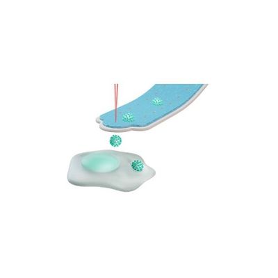
Precisely deposit a single virion
Single virions can be placed at an exact location on your cell of choice
Inject a single virion
Directly inject a single virion into the cytoplasm or the nucleus of a specific cell
Measure biophysical changes
Measure mass changes, variation in stiffness, and changes in adhesion force
Isolate, extract & analyze
Isolate infected cells - or take single cell biopsies - for further expansion or analysis
Observe & monitor
Continuously monitor the cell and observe the surrounding culture via integrated microscope and tracking software
FluidFM introduces new experimental possibilities to virology by allowing full control over how many virions interact with user-selected cells in adherent cell cultures. This promises new insights into:
- Cell entry and infection mechanism
- Cellular response, virus cooperativity and virus life-cycle stages
- Proliferation, spreading rate and cell to cell infection
By placing defined numbers of virions onto a cell, the host cell defense against the virus can be quantified. Therefore, infection probability, the host defense limitations, and the cooperation between virions can be studied.
Monitor cell-to-cell spread
Thanks to the integrated CO2- and temperature incubation unit as well as the epi-fluorescence microscope, FluidFM operates in a fully cell culture-compatible environment. This preserves the cell culture context of the infected cells and, together with the software-supported automated tracking function, allows long-time observation of the infected or manipulated cell. This allows following in detail how the viral infection spreads from the host cell to its neighbors and onto the rest of the culture.
Gently pick up a single cell from adherent or suspension culture and place it into another well plate with micrometer precision, fully preserving the cells’ viability. This enables to study the effect of introducing single infected cells into a healthy culture. The same method can also be used to place healthy cells, resistant cells, or drug-treated cells onto infected cultures.
Isolate cells of interest for further expansion or analysis
Single cells can be isolated from a culture based on morphology or fluorescent markers. Staying fully viable, these cells of interest can then be expanded in a new dish or processed for further proteomic or transcriptomic analysis. It is even possible to take single cell biopsies for analysis.
The integrated force feedback of FluidFM Probes allows quantifying mechanical interactions with pN force resolution. Measure biophysical changes induced by the infection of single cells, such as variations in stiffness, changes in adhesion force and, depending on the system, even changes in mass. FluidFM thus allows to correlate morphological with mechanical changes induced by the virus onto its host cells.