

Robotic-Assisted Bronchoscopy System for Lung Cancer Statistics - Medical / Health Care
Lung cancer is the leading cause of cancer deaths worldwide. More Americans die each year from lung cancer than from prostate, breast and colon cancers combined.1
Lung cancer has the lowest 5-year survival rate (18%) when compared to other common cancers. This is in part because most cases are not diagnosed until a later stage. However, if diagnosed and treated early, survival rates increase to 55%.2
One reason lung cancer is so deadly is that detection, diagnostic and treatment processes are ineffective.
In early stage lung cancer, suspicious lung nodules tend to be very small and deep in the periphery of the lung. This makes it difficult for physicians to safely and reliably diagnose them. Although there are a variety of diagnostic options currently available to biopsy lung nodules, all have limitations in accuracy, safety or invasiveness.
Surgical excision of a suspicious lung nodule is the most invasive option, has the highest risk to the patient and generally requires a stay in the hospital.
A transthoracic needle biopsy (TTNA) is less invasive, but it requires a needle to be inserted through the chest wall from outside the body. This can result in a collapsed lung (pneumothorax) up to 42% of the time, with 17% of patients requiring a chest tube to re-inflate their lung.3
Traditional flexible bronchoscopy or transbronchial needle aspiration (TBNA) are the safest of the diagnostic procedures. A flexible bronchoscope is often used successfully when diagnosing lung lesions, but they are limited in their reach and often fail to access the outer areas of the lungs. Unfortunately, up to two-thirds of all lung nodules are located in the periphery and may be unreachable using a standard bronchoscope.4
To aid bronchoscopy in the diagnosis of small, peripheral nodules, adjunct technologies such as radial ultrasound probes and electromagnetic navigation systems have been introduced. A recent multi-center registry with data from 15 centers and 581 patients demonstrated that these technologies have only achieved a diagnostic yield of 53.7% when they are used alone or combined.5
Auris is committed to providing physicians with safe and reliable technology to diagnose patients with small, peripheral nodules.
Current bronchoscopic technologies have limitations when it comes to reliably accessing and targeting peripheral lung nodules, resulting in a lower than desired diagnostic yield and possibly subjecting patients to more invasive diagnostic interventions with higher complication rates.8, 9, 10
When physicians diagnose a small nodule deep in the lung, they must be able to:
- drive the bronchoscope far enough to reach the nodule
- control the biopsy tools for precise and accurate tissue collection for diagnosis
Manual bronchoscopes have two-directional steering, limiting a physician`s ability to advance the scope through tortuous anatomy. To overcome this limitation, physicians must use an expensive sheath or catheter to extend their reach beyond the scope. Often this is combined with electromagnetic navigation to help guide the sheath to the area of the nodule. However, these tools do not provide any direct visualization, potentially contributing to the likelihood of a non-diagnostic procedure.
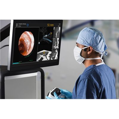
The MONARCH® Platform improves upon existing minimally invasive techniques by integrating robotics, micro-instrumentation, endoscope design and data science into one platform to empower physicians.
“The MONARCH® Platform was designed to allow physicians to diagnose small, peripheral lung nodules with greater precision than ever before,” said Eric Davidson, President, Flexible Robotics at Auris Health.