

- Home
- Companies
- Firstkind Ltd
- Applications
- Wound Therapy Applications Device for ...
Wound Therapy Applications Device for Mixed Aetiology Leg Ulcers - Medical / Health Care
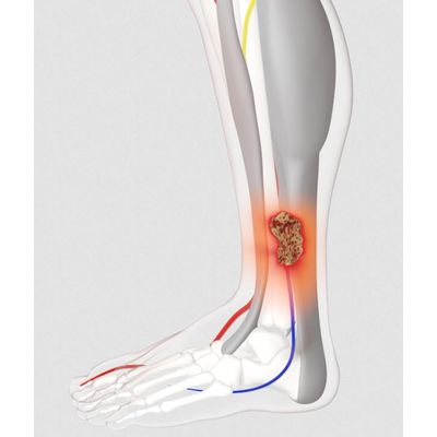
Leg ulcers occur as a result of various aetiologies. It is thought that more than half are of venous origin, while the remainder are due to arterial, mixed arterial/venous or other pathologies.
The prevalence of mixed aetiology leg ulceration is likely to increase as the population becomes older, as elderly people are more likely to have arterial disease2.
Accurate assessment of venous and arterial function of the leg is therefore paramount in determining the treatment of leg ulcers3.
The cause:
- Wounds, including mixed arterial ulcers often have impaired blood flow4,5.
- Impaired calf muscle pump function increases venous stasis and venous hypertension, and can negatively impact the severity of venous ulcerations6,7,8,9.
The treatment:
- Improved blood circulation results in enhanced wound closure5,10 a natural healing response.
- The geko™ device increases venous, arterial and microcirculatory blood flow in the lower limb in patients with chronic venous insufficiency11,12 and intermittent claudication13. It also reduces oedema14,15, augments the calf muscle pump16 and maintains TCpO2 – promoting conditions suitable for wound healing17,18,19.
- Where an ulcer has an arterial component to its aetiology; the gekoTM device is more efficacious than compression therapy, by virtue of the augmentation of arterial inflow18. Additionally, since healing any ulcer requires perfusion at the wound bed, the augmentation of microcirculatory flow brought about by the gekoTM device can be expected to be beneficial.
A targeted approach to the healing of complex wounds
A case series evaluation conducted by Professor Keith Harding, at the Welsh Wound Innovation Centre (WWIC) in Cardiff, has investigated the therapeutic effect of the geko™ device on wound healing outcomes over an 8-week period.
The case series used the geko™ device in patients with mixed aetiology leg ulceration in conjunction with appropriate or tolerable compression therapy and in arterial ulcers as a standalone therapy prior to or after surgery.
The research findings support use of the geko™ device in patients with painful venous and mixed leg ulceration in conjunction with best practice standard care. The geko™ device was effective in reducing the wound surface area and increasing the mean percentage of granulation tissue formation. 52% reported a substantial reduction in wound pain 20.
Reported as easy to use, the geko™ device facilitates self-management and empowered and engages patients and their carer’s in their care20.
Contrast speckle imaging data from a single WWIC case study (shown above) demonstrates a 225% increase in microcirculatory blood flow in the wound bed, and a 67% increase in microcirculatory blood flow surrounding the peri-wound area, after activation of the geko™ device. The increase in blood flow to the wound bed promotes conditions favourable to wound healing20.
The geko™ device is an adjunctive therapy
Consider the geko™ device for any or all of the following when:
- A wound has healed less than 30% in 4 weeks of care.
- Patient has a fixed ankle joint or poor ankle flexion.
- Lower limb muscle pumps are impaired or inactive.
- Oedema management has been a factor in wound healing.
- Wound, ischemia and edema may be the underlying cause of pain.
- Patient has an increased risk of Venous Thromboembolism.
- Compression cannot be tolerated or is contraindicated (use of the geko™ device has been shown to enable patients to tolerate compression who have not tolerated it before).
Health Economics
The geko™ device can contribute to a reduction in the costs of unnecessary call out for community nursing teams or unscheduled visits to wound clinics. Patients are enabled to self-manage the geko™ device to take control over their symptoms whilst resuming their normal daily activities.