

- Home
- Companies
- Artel, Inc. by Advanced Instruments
- Articles
- Imaging Complex Cell Morphology: ...
Imaging Complex Cell Morphology: Mesenchymal Stem Cells (MSCs)
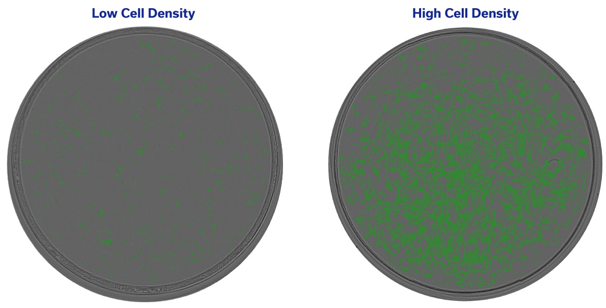
MSCs exhibit specific morphology, which makes them unique compared to other cell types typically used in CLD. While forming spindle-shaped, elongated, and transparent cell projections, the MSCs can pose a significant challenge when it comes to imaging and subsequent image analysis.
Many imaging systems on the market are simply not fit for precise detection of these unusual cell types. Systems with poor image quality and low contrast tend to struggle to distinguish MSCs from the background or any surrounding artifacts. This can introduce a lot of inaccuracies during image analysis especially when working with a range of cell densities – an important step included in clonal outgrowth quantification, also often referred to as confluence assessment.
Regular checks of cell confluence during clonal outgrowth are necessary for the determination of the best scale-up conditions, clone ranking, and overall standardization of culturing conditions across samples and projects.
Manual image analysis tends to be subjective and time-consuming. Frequently the results are also biased depending on the level of the operator’s experience. Automation of image analysis is an important step in the process, which not only eliminates subjectivity but also introducing consistency and time-savings.
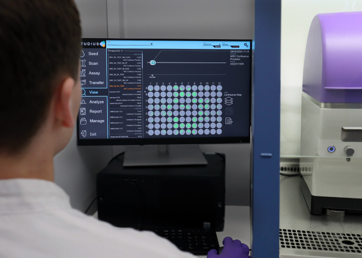
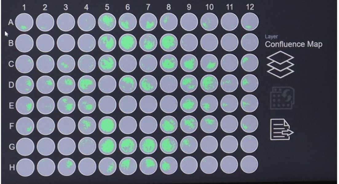
VIPS® PRO and Cell Metric® X both can precisely measure the confluence of MSC-like cell types using a new Artificial Intelligence (AI)-driven algorithm developed specifically for cells with elongated, spindle-like morphology.
In addition to an extremely accurate MSC confluence readout, the scan delivers three image planes for each well allowing the end user to make comparisons at lower densities and decide which image plane to use in the Clonality Report. All raw images collected using STUDIUS™ software are easily exportable for any further analysis.
- Application: Confluence of MSC and MSC-like cell types.
- Plate Type: Validated 96-well plates
- Validated 384-well plates
- Instruments: VIPS PRO Single Cell Seeder
- Cell Metric X Clone Imager
- Scan Times: In line with Confluence Precision Scan
- Number of Image Planes: 3 image planes
- Part Number: CC-4400