Fluorescence Imaging Articles & Analysis: Older
10 articles found
PEG-modified nanoparticles can be used as biomarkers for detecting and imaging biomolecules. Carboxylic acid PEGs can be combined with markers such as fluorescent dyes or radionuclides to achieve high-resolution imaging and fluorescent labeling in vivo. In addition, carboxylic acid PEGs can be used to construct ...
Copper-Free Click Chemistry for Clickable Drug Surrogates Copper-free click chemistry has presented intriguing opportunities for the development of clickable drug surrogates, particularly in the context of fluorescent imaging of target of interest (TOI) proteins within live cells. ...
These dyes emit light when excited by specific wavelengths, allowing researchers to visualize the liposomes in real-time using fluorescence microscopy or other imaging techniques. Benefits of fluorescent liposomes Real-time tracking: Researchers can observe the movement of liposomes within tissues and organs, understanding their uptake by ...
Assay for transposase-accessible chromatin using sequencing (ATAC-seq) was developed and was further applied to single cells9,10. Imaging chromatin accessibility in fixed cells using fluorescence-labelled DNA oligomers assembled in Tn5 (ATACsee)11 suggests that it is feasible to profile chromatin accessibility in situ. ...
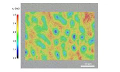
Welcome to Edinburgh Instruments newest blog celebrating our work in Raman, Photoluminescence, and Fluorescence Lifetime Imaging. Every month we will highlight our pick for Map of the Month to show how our spectrometers can be used to reveal all the hidden secrets in your samples. ...
Fluorescent dye-labeled peptides can be visualized by fluorescence microscopy or other fluorescence visualization techniques. ...
Title: Detection of Thyroid Cancer and Central Lymph Node Metastases using c-Met Enhanced Optical Imaging Objective: The main objective of this phase I pilot study is to assess the feasibility of EMI-137 for the intraoperative detection of thyroid cancer and central lymph node metastases in PTC using optical fluorescent imaging Status: Study ...
Fluorescence in situ hybridization (FISH) is a technique that prepares acceptable results for molecular imaging biomarkers to precisely and dependably detect and diagnose disorders which are sign of cancers. ...
Early attempts were all based on multiplexed single-molecule fluorescent in situhybridization (smFISH) via spectral barcoding and/or sequential imaging ( Pichon et al., 2018 ; Trcek et al., 2017 ). ...
Quantum dots have the potential to be used as contrast agents for molecular imaging in vivo, but there are many challenges in optimising such procedures, both in pre-clinical animal models and in the potential clinical applications. ...


