

- Home
- Companies
- Diagnosys LLC
- Articles
- Visual electrophysiology for infants ...
Visual electrophysiology for infants and young children continues to evolve
Poor fixation and tracking behaviors in infants and young children can arise from a wide range of underlying causes. Imaging modalities such as fundus photography, fundus autofluorescence, fluorescein angiography, and optical coherence tomography are available but typically require anesthesia. The saccadic vector optokinetic perimeter (SVOP), with its built-in eye-tracking, was designed to substitute for standard visual field testing in young children, but it still requires a degree of cooperation and fixation.
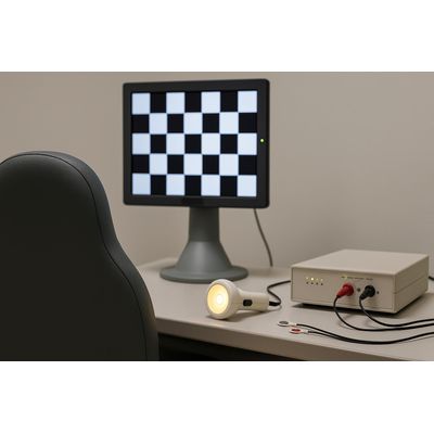
Visual electrophysiology tests designed for pediatric patients do not require anesthesia or pupil dilation. Tests based on flash stimulation do not require fixation. These tests provide objective assessments of the retinal photoreceptors and the entire visual pathway up to the visual cortex. More specifically, a combined recording of electroretinography (ERG) and visual-evoked potentials (VEP) can functionally dissect the visual pathway and identify the site of dysfunction in pediatric patients, whether retina, optic nerve, or visual cortex is the primary cause of vision loss.
Modern visual electrophysiology systems allow ERGs and VEPs to be recorded simultaneously, which is convenient when testing infants and young children. Once electrodes are in place, flash and pattern tests can be performed sequentially within a single session. In a webinar hosted by Diagnosys, Dr. Oliver Marmoy from Great Ormond Street Hospital (GOSH) demonstrated that a full suite of tests, including electrode placement, can be performed on a young child in under 30 minutes.
Flash ERG adaptations for young children
In the early 1990s, GOSH developed an alternative protocol for electroretinography (ERG) testing in children, now widely used internationally. This approach categorizes retinal abnormalities into the rod system, the rod photoreceptor, the cone system, and those affecting the inner retina, as reflected in the ERG waveform components.
A key aspect is that testing requires only 5–10 minutes, with natural pupils and minimal dark adaptation. It uses skin electrodes, which are safe for infants and young children. It also delivers handheld flash stimulation from a remote distance to minimize intimidation and enable testing in alert children of all ages. For infants and young children, interaction and play, with cartoons, music, and sounds, are used to garner attention and gaze for successful recording. The ColorFlash stimulator is optimized for testing children with short attention spans and features built-in animal sounds to aid engagement.
During a 30-minute session, clinicians can engage the child with cartoons, play, and treats. In pattern-stimulus testing, clinicians may alternate between the stimulus test and engaging visuals to help the child fixate on the screen.
Glasgow sweep VEP
The Glasgow sweep Visually Evoked Potentials (sweep VEP) is an ISCEV extended protocol for objectively assessing visual acuity. It is designed for children and patients who have difficulty maintaining fixation. The method uses a thresholding algorithm that automatically adjusts the checkerboard size of the pattern VEP until the final threshold is reached. The spatial frequency limit, determining visual acuity, is based on the final step identified by the algorithm, enabling acuity assessment from 20/20 to 20/2000.
Pattern stimulus adaptations for young children
Clinical researchers in the Netherlands developed a method of superimposing an animated cartoon on a pattern stimulus. The cartoon, La Linea, depicts a single-line, talking figure with music in the background. This minimalist cartoon is optimized to minimize interference with the pattern stimulus. Although tested in adults, the study recorded gaze movements across fixation to saccades. Findings showed that, in pattern VEP, the P100 amplitude and peak time were reduced with the cartoon present compared to a standard fixation-point test. While the reduction was statistically significant, it remained within clinically acceptable limits, suggesting the cartoon superimposed on the pattern stimulus can be suitable for testing young subjects.
Pattern VEPs for gene therapy monitoring in young children
The ophthalmology field views FDA approval of Luxturna in 2017 and EMA approval in 2018 as landmark milestones for retinal gene therapy. Diagnostic methods that supported Luxturna’s drug trials included full-field stimulus testing (FST) and multiluminance mobility tests (MLMT). However, both require patient participation, presenting challenges with young children. Pattern VEPs offer minimal cooperation and provide objective measures of the visual cortex. This method was evaluated as an alternative to FST and MLMT for managing young children treated with Luxturna. A retrospective study of 27 eyes in Luxturna-treated children from 2020 to 2023, conducted by researchers affiliated with Great Ormond Street Hospital, University College London, and The Tony Kriss Visual Electrophysiology Unit, reported that pattern VEPs were reproducible and showed increased amplitude after treatment.
An industry committed to advancing pediatric ophthalmology
Children who are too young to communicate their visual health are particularly vulnerable. Visual electrophysiology offers objective modalities adapted for young children. Modern protocols do not require anesthesia or mydriasis. Clinicians have demonstrated the practicality of these protocols in diagnosing and managing very young patients. Ongoing visual electrophysiology studies focused on infants and young children continue to be published, contributing to the knowledge base. The visual electrophysiology community remains dedicated to advancing pediatric ophthalmology.
Original: https://www.diagnosysllc.com/visual-electrophysiology-for-infants-and-young-children-continues-to-evolve/