

- Home
- Companies
- JPK BioAFM Business - Bruker Nano GmbH
- Products
- CellHesion - Model 200 - Cell/tissue ...
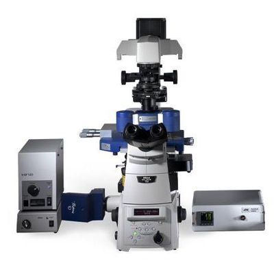
CellHesion - Model 200 -Cell/tissue Mechanics and Cell Adhesion
The CellHesion® 200 is an integrated system for measuring cell-cell and cell-substrate interactions. The system can also be used to quantify cell elasticity and cellular response to external mechanical stress or can map tissues.
The quality of the force spectroscopy data is vital for successful analysis. The lowest electronic noise floor and the most rigid mechanical design are essential. The highest accuracy and stability of the instrument is ensured by integrated capacitive position sensors with exceptional performance. For minimized drift, the system design is symmetric.
For the detection of smallest variations in force curves JPK improved the sensitivity and increased the detection bandwidth without compromising the noise levels. The high sampling rate and virtually unlimited number of data points per force curve complete this unique instrument.
- A single living cell is chemically bound to the cantilever sensor (e.g., through a fibronectin coating) under optical control.
- This cell is brought with a defined force into contact with the binding target (molecular layer, implant surface, single cell, confluent monolayer) on the substrate (slide, coverslip or Petri dish).
- After a user-defined reaction time the cell on the cantilever is separated from the substrate cell by retracting the cantilever in vertical direction (z-axis) through a piezo actuator.
- The cell resists the attempt of removing it from the surface if it adheres to the target. Therefore the cantilever bends noticeably, which is measured by a detector.
Because, in physical terms, a cantilever is a leaf spring, the actual adhesive forces and energies can be derived from the measured bending. This allows the identification of single-molecule binding events that contribute to the adhesion. The experiment is repeated many times with the same cell, with different cells, on different targets and with different conditions to gain statistically relevant information.
The result of a single measurement cycle is a force vs. distance curve, which allows to determine single molecule events, the “work of removal” W, tether formation, the maximum adhesion force and viscoelastic parameters.
The easy and complete integration of the CellHesion® 200 into inverted research microscopes from leading manufacturers such as Carl Zeiss, Leica, Nikon and Olympus makes it a powerful combination. Techniques such as epifluorescence, confocal microscopy or Superresolution microscopy (STED, STORM/PALM) can be used simultaneously with the CellHesion® system.
Observation of target cells with fluorescence techniques such as TIRF, CLSM, FRAP or Ca2+ imaging parallel to CellHesion® experiments gives insight about the molecular mechanisms involved in adhesion processes or cytoskeleton dynamics. All modes of optical transmission illumination techniques such as DIC or phase contrast can be used simultaneously. This is an important feature when transferring cells to the force sensor (cantilever) or to check the condition of the specimen. Structural information derived by these optical methods can be overlaid with functional data that is determined by force measurements via the DirectOverlay™ software feature, when the CellHesion®200 is used together with the motorized precision stage or the precision mapping stage add-ons.
CellHesion® 200 is tailor-made for the specialized requirements of working with living cells. The use of standard substrates such as 35mm Petri dishes with or without glass bottom or round coverslips makes cell cultivation and handling easy.
Temperature control from 15°C to 60 °C, fluid exchange and ports for CO2 control are integrated in the JPK PetriDishHeater™ or in the JPK BioCell™ designed for coverslips. As an option, the CellHesion® 200 can be integrated in existing incubators (ask for model).
All parts of the setup which are in contact with the sample can be sterilized. CellHesion® 200 offers enough vertical-axis travel range to handle large cells and separate even well-adhering cells from their substrates.
- Stiffness and elasticity mapping from single cells to substrates and tissues
- Cell-cell and cell-substrate interactions
- Cell adhesion and tether formation
- Automated mapping of sample properties over a large range for structured substrates, microspheres, cells. etc. with the new HybridStage™
- Biomaterial studies, biofouling, biosensors, capsules
- Implants coatings and cellular biochips
- Applications in microbiology and virus research
- Pharmaceutical studies such as drug delivery mechanisms
- Applications in food, paper or textile industry on fibers, coatings or powders in air or liquid
- Binding studies such as receptor/ligand or antibody/antigene
- Testing functionalized surfaces
- Innovative platform for cell or tissue mechanics and adhesion experiments
- Characterization of cell/cell or cell/substrate interaction, cell elasticity, tether formation, adhesion, and cellular response
- Quantitative measurements from single molecules to entire cells and tissue, organelles and embryos with highest precision
- Automated mapping of sample properties over a large range for structured substrates, microspheres, cells. etc. with the new HybridStage™
- Intuitive user interface with ExperimentPlanner™ for perfect workflow
- Integrates with advanced optical imaging such as DIC, phase contrast, fluorescence, confocal microscopy, Superresolution, TIRF, FRET, FLIM, etc.
- Widest range of modes and accessories
Bruker’s BioAFMs allow life science and biophysics researchers to further their investigations in the fields of cell mechanics and adhesion, mechanobiology, cell-cell and cell-surface interactions, cell dynamics, and cell morphology. We have collected a gallery of images demonstrating a few of these applications.
- Innovative platform for cell adhesion/cell mechanics research for measurements from single-molecules to entire cells and tissue
- Compatible with inverted microscopes for combined experiments, e.g., Zeiss Axio Observer, Axiovert A1, Nikon Ti/TE 2000, Nikon Ti2, Olympus IX series and Leica DMi series
- Compatible with all major light microscopy techniques such as DIC and phase contrast or fluorescence and confocal techniques such as TIRF, FRAP, LSM and others
- Compatible with upright macroscopes and stereo microscopes such as Zeiss AxioZoom V.16, Leica Z16 ApoA, Leica M205FA and Olympus MVX10
- Automated range adaption in z direction for strong corrugated surfaces
- Cantilever sensor lifting system with >110µm travel range with closed-loop control through high-speed capacitive sensor feedback
- Equipped with motorized precision stage, in particular for tissue probing, within 20×20mm range
- Living cell studies in native environment with tempeature control, perfusion and gas flow (CO2) on coverslips with the JPK BioCell™ or 35mm Petri dishes with or without glass bottom from Wilco, BD, WPI with the JPK PetriDishHeater™
- All parts which came in contact with the sample are easy to clean
- Compatible with standard incubators (specify model)
- High-throughput determination of cellular interaction parameters through JPK software batch processing
- Single-molecule events, work of removal, maximum adhesion forces, number of unbinding events and viscoelastic parameters such as Young’s modulus in one system