

- Home
- Companies
- Kowa American Corporation
- Products
- Kowa - Model VX-20 - Combination ...
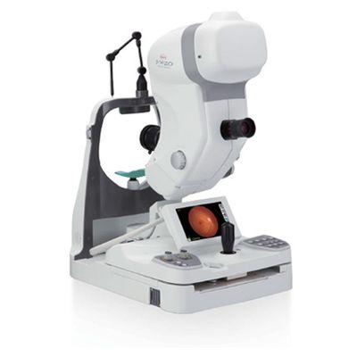
Kowa - Model VX-20 -Combination Mydriatic/Non-Mydriatic Retinal Camera
The VX-20 is Kowa`s unique, hybrid combination retinal camera that offers versatile, high quality retinal imaging in one easy-to-use system. Due to its outstanding features, the VX-20 remains the first choice for clinicians. With a built in PC and high-resolution camera, this fundus camera is a versatile diagnostic tool with stand-alone capability. The VX-20 fits perfectly within the clinical environment. The VX-20 offers enhanced functionality at the touch of a button allowing the clinician to switch quickly between mydriatic & non-mydriatic modes. It includes a 7 inch wide, LCD color touchscreen that is simple to use and ideally positioned to ensure easy patient observation. What`s more, the control panel`s updated design improves the operator experience, ensuring everything is within easy reach for the clinician.
5 Photography Modes Including FAF*
In addition to non-mydriatic color, mydriatic color, fluorescein angiography (FA), Red-free (RF), fundus autofluorescence (FAF*) is added to allow 5 photography modes.
75mm reduced height of examined eye from our previous model ("VX-10" Series) allows for photography in a more relaxed posture.
- Change the field of view with a quick push of a button
- Mydriatic mode: 50° or 30° field of view
- Non-mydriatic mode: 45° or 27° field of view
Through the Optical Viewfinder
In mydriatic mode, the fundus can be observed through the optical viewfinder, allowing image capture exactly as viewed. This feature is particularly helpful for shooting the peripheral part of the retina.
- Eye level indicator can be easily seen even in a darkroom
- Stable chassis makes it easy to assist in patient eyelid opening
- Expanded downward tilt angle up to 11° facilitates upward-angle
shots which have been difficult in conventional models
*As compared to previous models
- Special Built-In Digital Camera
- Wide-screen 7" touch LCD monitor
- Icons strategically placed for improved operator experience
- Available buttons are illuminated in chosen photography mode to enable smooth and quick photography
- Adjust the chin rest up/down or switch the field of view with the simple push of a button
- φ3.5mm or φ4.0mm options in Mydriatic mode
*The SP mode is not supported in RF mode.
*With φ3.5mm photography, some eyes may cause flare around their circumference.
- Central/Disc/Macula/Peripheral and external fixation light can be switched with a single push of a button
- Adjustable photographic ISO sensitivity, light intensity, and diaphragm
- Photographic settings can be easily adjusted. To allow patient friendly photography or to compensate for insufficient light intensity in later FA phases, the ISO sensitivity can be increased or decreased.
- To reduce the photophobia of the patient, photography in mydriatic color, FA, RF, and FAF modes under monitor observation is possible
Working Distance: 39mm (Between the examined eye and the objective lens)
Monitor: 7 Inch Widescreen Touch LCD
Dimensions: 390 (W) x 540 (D) x 720 (H) mm
Weight: 39kg/86lbs
Working Distance Adjustment: Luminous Dot Indication (ON/OFF Switch)
Internal Fixation Target: Central, Disc, Macula, Peripheral
External Fixation Target: Red/Green LED
Camera: 24 Megapixel CMOS Sensor
Photography Modes: Non-mydriatic Color, Mydriatic Color, Fluorescein Angiography (FA), Red-Free (RF), and Fundus Autofluorescence (FAF)
Field of View: Non-mydriatic Mode: 45°/27° Mydriatic Mode: 50°/30° (SP: 45°/27°)
Minimum Pupil Diameter: Non-mydriatic Mode: ⌀4.0mm (SP⌀3.5mm) Mydriatic Mode: ⌀5.5mm (SP1 ⌀4.0mm/SP2 ⌀ 3.5mm)
Focusing: Split Luminous Bars Coincidence
Range of Focus Adjustment for Compensation of Patient`s Refracti: Without Compensation: -12D to +13D -Compensation: -32D to -10D +Compensation: +10D to +35D
Range of Diopter Adjustment of the Optical Finder: -8D to +5D
Light Source: For Observation: Halogen Lamp For Photography: Xenon Flash Lamp
Flash Compensation: ±5 Steps
Exposure: Appropriate exposure is automatically set based on the 37 steps (0.6WS to 300 WS) of field angle, filter, and photography mode
Adjustment Range: Forward/Backward: (gross) 90mm, (micromotion) approximately 17.5mm Left/RightL (gross) 140mm, (micromotion) approximately 17.5mm Up/Down: 30mm Tilting:(Elevation) 11°, (Depression) 15° Panning: (left/right): 30° horizontally
Recording Medium: CF Memory Card
Interface: USB: VK-2 Connection, Printer Connection, Card Reader Connection LAN: Image Output
Power Saving Function: Yes
Power Supply: Input: AC 120/230V 50/60Hz Power Consumption: 250VA 1500VA (Max)