

- Home
- Companies
- Leica Microsystems GmbH - a Division of ...
- Products
- Leica - Model ARveo 8 - Digital ...
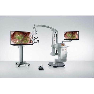
Leica - Model ARveo 8 -Digital Visualization Microscope for Neurosurgery
ARveo 8 unites information from AR fluorescence, IGS systems, and endoscopic image feeds providing an enhanced visualization for more informed and precise neurosurgery.
With ultra-fast processing and an intuitive graphical user interface the ARveo 8 digital visualization microscope helps to enhance efficiency across the entire team.
Leica Microsystems` EnhancePath concept for future upgradeability and system compatibility of the ARveo 8 provides a seamless way to evolve into the digital future of neurosurgery, smoothly and confidently.
Enhanced visualization for greater precision
- ARveo 8 enhances visualization by adding layers of information to the microscope image so that neurosurgeons can obtain more information while operating on a patient.
- ARveo 8 combines world-renowned optics with the ability to combine pre- and intraoperative imaging and augmented reality fluorescence, so that neurosurgeons can make precise, confident decisions.
- Ultra-fast processing in the ARveo 8 reduces latency by 44%* and delivers information to the surgeon faster—further amplifying precision.
- ARveo 8 enables surgeons to truly work “in the now” with greater confidence for better patient outcomes.
Augmented vascular surgery
The GLOW augmented reality (AR) ecosystem is an integral part of the ARveo 8 digital visualization capabilities.
A sophisticated imaging sensor and algorithms capture, optimize, and combine multiple spectral bands of visible and fluorescent light.
The result is a fully synchronized, real-time, augmented view of the surgical field.
GLOW800 augmented reality (AR) fluorescence and ICG enable you to observe cerebral anatomy in natural color, augmented by real-time vascular flow, with full depth perception.
Anatomy and blood flow in one image
When combined with ICG, GLOW800 augmented reality (AR) fluorescence allows you to observe cerebral anatomy and blood flow in white light.
Forget yesterday`s need to recall and reconcile black and white NIR blood flow video with the natural anatomical view—enjoy one single view of anatomy and blood flow.
- Stay orientated thanks to full depth perception without dark peripheries through image homogenization
- Work confidently in GLOW800 mode for removal of AVMs, clipping of aneurysms, performance of a bypass or a microvascular decompression
The augmented image is displayed in the oculars via CaptiView image injection and on the monitors in the OR.