

Circle Cardiovascular Imaging Inc.
- Home
- Companies
- Circle Cardiovascular Imaging Inc.
- Products
- Model ADAS 3D - Fibrosis Imaging for ...
Model ADAS 3D - Fibrosis Imaging for the EP Lab
ADAS 3D enables non-invasive pre-procedural planning for electrophysiologists. cvi42 now integrates ADAS 3D – a solution that enables pre-procedural planning by incorporating non-invasive MR/CT imaging to quantify LV/LA fibrosis and LV wall thickness and display surrounding anatomical structures.
Most popular related searches
ADAS 3D LV helps visualize and quantify LV fibrosis (MR) and helps visualize surround anatomical structures (CT).
- Visualize the fibrosis (LGE-MRI) in 3D colored images
- Quantify Core scar and Border Zone (BZ) volumes
- Navigate 9 layers from endo to epicardium
- Visualize auto detected corridors of BZ tissue
- Quantify LV wall thickness (CT)
ADAS 3D LA helps visualize and quantify LA fibrosis (MR) and helps visualize surrounding anatomical structures (CT).
Highlights
- Visualize the distribution and quantify the amount of enhanced fibrosis
- Visualize and navigate around the LA in 3D
- Display adjacent structures, including the esophagus
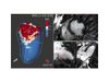
- ADAS 3D LV image synchronized with DICOM images
- Colored lines in DICOM image represent the areas in the ADAS 3D image for that slice
- 3D volume of Core Scar (CS) is shown on all layers from 10% (endocardial) to 90% (epicardial)
- CS is red, BZ is green-yellow, healthy tissue (HT) is blue
- Corridors of BZ tissue in the 10, 20, 30, 40% layers
- The centerline of the BZ is illustrated with the solid white line
- White silhouette of epicardial surface
- Right Ventricle shell illustrated in green