

- Home
- Companies
- ACIST Medical Systems
- Products
- ACIST - Model HDi - High-Definition ...

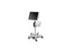
ACIST - Model HDi -High-Definition IVUS System
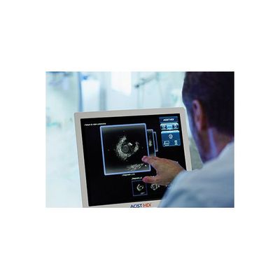
ACIST HDi High-Definition IVUS System enables optimized visualization to inform and illuminate coronary and peripheral intervention strategy through advanced imaging modes, improved deliverability of the Kodama IVUS Catheter and interactive compact console.
- HDi with enhanced imaging modes provides a better defined MJS image for preprocedural planning and post procedural assessment1
- Interactive compact console with touch screen for rapid analysis and small footprint for easy cath lab integration
- Improved deliverability2 and optimized imaging with the offset distal tip and unique variable flexibility (VariRex) imaging window of the Kodama HD IVUS Catheter
LumenView
Darkens the coronary lumen for better border detection.
SilkView
Increases gray scale for finer blood speckle, tissue and plaque differentiation.
ClassicView
Optimizes the balance of high resolution and depth of penetration and enables full vessel wall visualization.
Better visualization of media than OCT for optimizing stent sizing4
Enables enhanced imaging by providing sufficient penetration at 60 MHz to see the media layer, even in larger plaque volumes4, so the physicians can maximize the stenting cross sectional area and may lead to better patient outcomes5
IVUS Use changed the treatment strategy during the procedure 74% of the time6
IVUS-guided stents also had low proximal & distal plaque burden, & higher MSA
compared to the angio-guided group7
8x more detection of lipid pools at 60 MHz than 40 MHz8
IVUS-guided PCI is beneficial for preventing the Stent Edge Restenosis in cases with diffused plaque lesions compared to angiography alone9
IVUS is less expensive and more cost effective than
angiography in 71% of PCI procedures10
IVUS provides improved outcomes at lower costs, with greater economic benefit especially in higher risk subgroups (diabetes, renal insufficiency, ACS)