
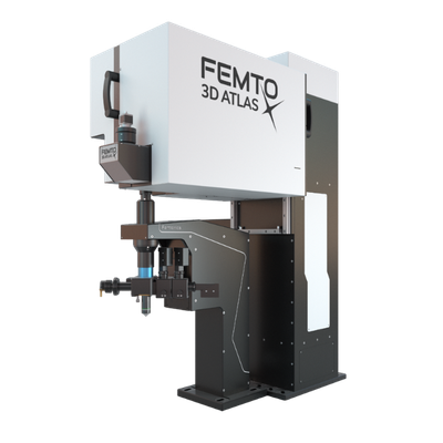
FEMTONICS - Model FEMTO3D Atlas -High-Speed 3D Imaging and Photostimulation System for Neuronal Activity
The FEMTO3D Atlas by Femtonics is a state-of-the-art multiphoton microscope designed for advanced in vivo 3D imaging and photostimulation, particularly in the field of neuroscience. Distinguished by its innovative integration of acousto-optic (AO) technology, the system excels in capturing high-resolution neuronal and dendritic activities with impressive speed and precision. Its capacity to perform simultaneous 3D photostimulation and imaging extends traditional two-photon microscopy, eliminating artifacts caused by physical activity such as breathing or heartbeat through advanced motion correction techniques. With scanning speeds reaching up to 100 kHz, the Atlas can engage with neural networks in real-time, offering unique insight into synaptic plasticity and memory formation. The introduction of a second laser line allows for concurrent optogenetic stimulations and uncaging. This versatile platform serves as an invaluable tool for research labs worldwide, aiding pivotal neuroscience inquiries and enabling high-impact publications.
NETWORK IMAGING
Neural networks consist of thousands of neurons dispersed in 3D space, often across many cortical layers of the brain tissue. 3D random-access scanning performed by AO technology combined with two-photon microscopy is the best choice to reveal the dynamics of the neural networks while processing information. It enables you to record neuronal activity from cell populations consisting of up to thousands of cells in vivo. The 3D anti-motion technology combined with new scanning methods united in FEMTO3D Atlas provides a flexible solution to gather motion-corrected data in large volumes at high framerates during behaviour. Learn more about Network imaging.
Watch the interview and find out how the FEMTO3D Atlas microscope contributed to the Losonczy Labs’ research published in Neuron.
DENDRITIC IMAGING
3D random-access point scanning extended by drifting the focal point along scanning elements with various shapes and sizes allows imaging without interruption at multiple dendritic branches. The sampling is continuous during the drift, so this scanning mode gives more detailed spatial resolution without changing the overall scanning time which reminds as high as during point scanning. As a result, the function of thin dendritic segments or even spines, single action potentials can be brought to light. See more Chiovini et. al, Neuron (2014).
BEHAVIORAL STUDIES
Our anti-motion technology enables cell activity to be captured while an animal is moving in virtual reality and performing tasks. To preserve signals while imaging, scanning points are extended to drifted lines which are precisely fitted to each other resulting in surface or volume elements enclosing the target object. These elements cover not only the pre-selected ROIs but also the neighboring areas giving an opportunity for motion correction by preserving all fluorescent information during motions and decreasing the artefacts by more than one order of magnitude in behaving animals.
OPTOGENETICS
Photostimulation techniques allow activating cells in a selective and precise manner, which makes them advantageous for various biological applications. Enhancing two-photon microscopy with this capability makes this technology well suited for studies ranging from synaptic plasticity to experiments investigating learning and memory in vivo in the brain tissue. To the FEMTO3D Atlas Dichro extension, a second laser can be coupled enabling users to perform optogenetic stimulation or uncaging with calcium imaging, simultaneously, even in different cell populations.