

Epithelix
Epithelix - Model MUCILAIR -In Vitro 3d Human Upper Airway Epithelium
FromEpithelix
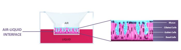
MucilAir is an in vitro cell model of the human airway epithelium cultured at the air liquid interface.
Most popular related searches
airway epithelium
metabolism activity
cytokine chemokine
metabolic activators
metabolic activities
metabolic activation
metabolic activity
pulmonary disease
allergy rhinitis
cystic fibrosis
Since 2006 we produce MucilAir™ with high standards of quality and reproducibility
- Reconstituted using human primary cells at low passage (P1)
- Remains fully differentiated and functional for over one year in culture
- Ready and easy to use
- Cultured in chemically defined serum-free medium
- Available worldwide in 24 well format
MUCILAIR™ IS AVAILABLE FROM SINGLE DONOR OR POOL OF DONORS
- Single donor or pool of donors available in nasal healthy version
- Different anatomical sites (Nasal, Tracheal or Bronchial)
- Patients with several pathologies:
- Healthy
- COPD (Chronic Obstructive Pulmonary Disease)
- Asthma
- Cystic Fibrosis (several mutations available)
- Allergic Rhinitis
- Smoker
MUCILAIR™ CHARACTERISTICS
- Air-liquid interface
- Production of mucus
- Active cilia-beating
- Active ion transport
- Tight junctions
- Metabolic activity
- Cytokines, chemokines, metalloproteinases release
MORPHOLOGY AND HISTOLOGY OF MUCILAIR™
- Radial cut of MucilAir™: C=Ciliated cells; M=Mucus cells; B=Basal cells.
- Cross section of cilia, the microtubules’ organization is visible.
- Tight junction between two cells of MucilAir™.
- Beginning of ciliogenesis.
CILIOGENESIS OF MUCILAIR™
A time course of cilia formation monitored by anti-β tubulin immunostaining over 60 days.
After 45 days the epithelia are fully differentiated and ciliogenesis is complete, the result can be observed by Electron Microscopy:
VIDEOMICROSCOPY OF MUCILAIR™
Videomicroscopy of a MucilAir™ epithelium reconstituted from a Healthy donor. Microbeads were applied on top of the epithelium, we can see an organization of the mucociliary clearance.