- Home
- Companies
- JenaValve Technology, Inc.
- Products
- JenaValve - Human Heart Anatomy Pump
JenaValve - Human Heart Anatomy Pump
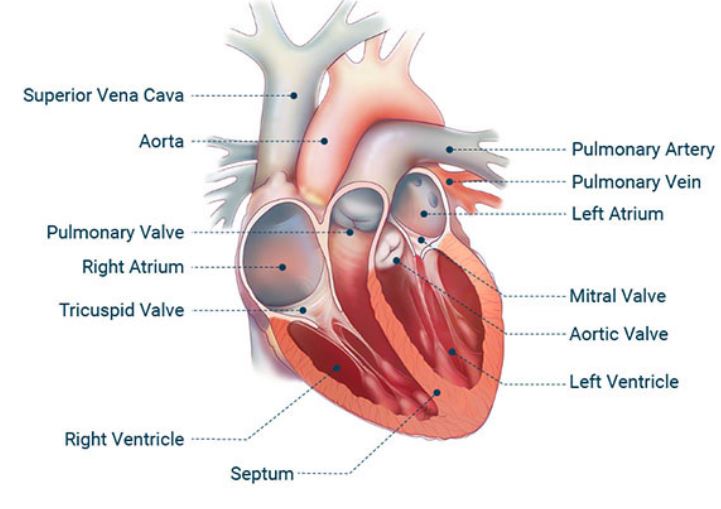
The human heart pumps blood into the arteries that carries oxygen and nutrients to all the tissues of the body. The heart is located in the center of the chest with its apex toward the left. It is the hardest working muscle in the body as it beats non-stop. If we want to understand how the heart performs its vital role, we will first have to look at its structure, i.e., cardiac anatomy.
The heart is a muscular pump with four chambers and an equal number of valves. The two chambers at the top of the heart are known as the atria, a right and a left. The two bottom chambers are the ventricles. The atria receive blood that returns from the different parts of the body, while the ventricles pump that blood back to all body tissues. Valves that separate the atria from the ventricles are called the atrioventricular valves. There are two: the tricuspid on the right and the mitral on the left. Valves at the ventricular outlets are called semilunar valves. The two semilunar valves are the pulmonary and the aortic.
The heart wall consists of three layers: the outer epicardium, the middle myocardium, and the inner endocardium. The epicardium and endocardium are thin layers. The myocardium forms the main bulk of the heart and is made up of cardiac muscle fibers. The outermost layer that surrounds the entire heart is called the pericardium. Vessels that carry blood away from the heart to the body are called arteries, while those that bring it back are called veins. The largest artery is named the aorta. It arises from the left ventricle.
The heart has an electrical system that originates and transmits cardiac impulses that cause the heart to beat. The system is made up of nodes and conducting fibers. If a person were to close their fist, slightly open it, and close it again, in rhythmic motion, this would be a simplistic display of the beating heart. The human heart is close to the human fist in size. It weighs around 8 to 10 ounces.
The events that take place in the heart between successive heartbeats constitute the cardiac cycle. Such events include the opening and closing of valves and contraction and relaxation of chambers. The cardiac cycle is split into two phases: systole and diastole. During systole, the ventricles contract and push blood into the arteries. During diastole, the ventricles relax and receive blood from the atria.
“Mayo Clinic.” Aortic Regurgitation Symptoms and Causes, Mayo Foundation for Medical Education and Research, 13 Mar. 2020, www.mayoclinic.org/.