

- Home
- Companies
- RayBiotech Life, Inc.
- Products
- Model L-Series - Label-Based Antibody ...
Model L-Series - Label-Based Antibody Arrays
Combining direct antigen-labeling technology with our vast library of array-validated antibodies, RayBiotech has created our largest antibody array to date: the Human L6000 Array. With the L-Series high density array platform, researchers can now detect up to 6000 proteins simultaneously, obtaining a broad, panoramic view of protein expression. Our newly expanded panel includes a wide variety of metabolic enzymes, structural proteins, epigenetic markers, neuroregulatory factors, in addition to our popular list of cytokines, growth factors, receptors, adipokines, proteases, and signaling proteins. Available on both glass slide and membrane formats, this array is ideally suited for biomarker discovery studies and exploratory screens.
Let Us Run L-Series For You
Ship us your samples (4 sample minimum) and our experienced service department will perform a high-density array of your choice. We can analyze up to 6000 human, 1308 mouse, or 1500 rat proteins and return to you a full data report. Click the button below to learn more.
- High detection sensitivity
- Easy to use; obtain results in 2 days
- No need for fractionation, depletion or extraction of samples
- Consumes only 20-30 ul of serum/plasma
- No dedicated equipment needed (glass slide or membrane)
- Accurate and reproducible
- As little as 59¢ per target
- High throughput expression profiling
- Discovering potential molecular targets for drug development
- Uncovering molecular mechanisms of drug action
- Identifying crucial factors involved in disease processes
- Discovering biomarkers for disease management
- Discovering expression patterns for molecular classification of diseases
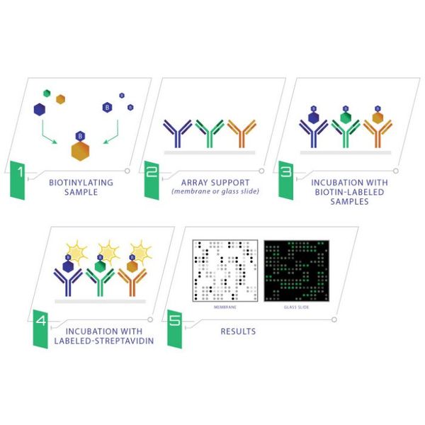
Through a simple labeling process, the sample proteins are directly conjugated to biotin, eliminating the need for a second antibody to develop the array signals. In this format, unintended antibody interactions are impossible, thus eliminating limitations on the size of the array panel.
- RayBio® L-Series Antibody Array Membranes or Glass Slides
- Spin Columns / Dialysis Tube
- Labeling Reagent
- Stop Solution
- Blocking Buffer
- Streptavidin-Conjugated HRP or Streptavidin-Conjugated Fluor
- Wash Buffer 1
- Wash Buffer 2
- Plastic Sheets
- Floating Dialysis Rack
- Plastic Incubation Tray
- Detection Buffer C
- Detection Buffer D
*Accessories include: 2-well or 4-well incubation chamber with gasket, protective cover, snap-on sides, adhesive film
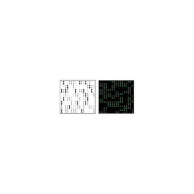
The cell culture supernates or serum is dialyzed before biotin-labeling step. Through a simple process, the primary amine of the proteins in the sample is biotinylated, followed by dialysis to remove free biotin. From here, the newly biotinylated sample is added onto the array membrane or glass slide and incubated at room temperature. After incubation with HRP-streptavidin or Fluorescent Dye-Strepavidin, the signals can be visualized either by chemiluminescence or fluorescence. The experimental procedure is simple and can be performed in any laboratory.