

- Home
- Companies
- SIFSOF LLC
- Products
- SIFSOF - Model SIFULTRAS-5.03 - Micro ...
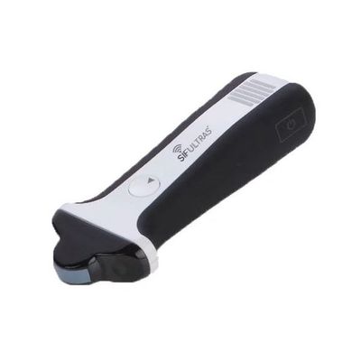
SIFSOF - Model SIFULTRAS-5.03 -Micro Convex Ultrasound Scanner Color Doppler Wireless
Color Doppler Wireless Ultrasound Scanner SIFULTRAS-5.03 has a Micro convex array Transducer. It has a Frequency bandwidth of 3.35 – 8.75 MHz and a Center Frequency of 6 MHz , a depth of 12.0 cm, a view field of 99°, a dimensions of 192mm x 72mm x 40 mm for applications such as : MSK, Pediatrics, Speech Therapy, Small parts, Abdominal, Bladder, Cardiac, Lung… and with only a 340 g weight. In addition, Color Micro-Convex Ultrasound Scanner SIFULTRAS-5.03 has b mode, m mode, color Doppler, power Doppler and pulsed-wave.
- Array Type: Micro Convex Ultrasound Scanner
- Frequency bandwidth: 3.35 – 8.75 MHz.
- Center Frequency: 6MHz.
- Field of view: 99°.
- Number of elements: 128.
- Channels: 32 Channels ADC System.
- Using Time: To 4.5 Hours.
- Weight: From 340g.
Color Doppler WiFi Ultrasound Scanner SIFULTRAS-5.03 is made for large calculation algorithm to create both B-mode and color mode. Furthermore, SIFULTRAS-5.03 has also a WiFi that connects to your mobile to the the images you’ve scanned.
Besides, Color Doppler WiFi Micro Convex Ultrasound Scanner SIFULTRAS-5.03 special development of US Image of Block Algorithm (UIBA, High-speed parallel image block algorithm), to demodulate high frequency, high contrast, high resolution and uniform of B-mode/Color image. SIFULTRAS-5.03 guarantee the high performance and reliable ultrasound imaging system solution.
Micro-Convex Ultrasound Scanner SIFULTRAS-5.03 has a various applications areas such as MSK, Pediatrics, Speech Therapy, Small parts, and it can be also applicable on Abdominal, Bladder, Cardiac, Lung…
- Give a visual overview of flow within the vessel or heart.
- Rapid identification of vessels, valves, turbulent flow.
- Evaluate flow direction and velocity.
- Measure volume and percent vascularity when combined with 3D Mode.
- Guidance for reproducible quantification of flow velocities using Pulsed-Wave Doppler.
- Locate area of stenosis or thrombosis.
- Determine the existence and amount of arterial plaques and associated turbulent flow.
- Find small vessels such as mouse coronary arteries, femoral and arcuate arteries.
- Evaluate blood flow after a stroke or other cases due to impaired blood flow.
- Observe blood flow to major organs such as heart, kidney, liver pancreas, carotid, abdominal aorta, and others.
- Wireless: Support IEEE 802.11 b/g Hands-Free Experience.
- Full Function: B Mode, M Mode, Color Doppler, Power Doppler and PW Are Ready.
- Endurance: More Than 4 Hours Continuously Use Experience.
- High Performance and Reliable Ultrasound Imaging System Solution.
- Large Calculation Algorithm To Create Both B-Mode and Color Mode.
- Special Development Of Ultrasound Image of Block Algorithm.
- Array Type: Micro Convex Ultrasound Scanner.
- Depth: 12.9 cm.
- Frequency bandwidth: 3.35 – 8.75 MHz.
- Center Frequency: 6MHz.
- Field of view: 99°.
- Number of elements: 128.
- Channels: 32 Channels ADC System.
- Using Time: To 4.5 Hours.
- Dimensions : 192mm x 72mm x 40 mm.
- Show images on your phone via Wi-Fi.
- 12 bit A/D converters with sample rate 50MHz to get the best image.
SIFULTRAS-5.03 can be applicable on Abdominal, Lung, OB/Gun, Fast, DVT and it can be also applicable on MSK, Pediatrics, Speech Therapy, Small parts, and it can be also applicable on Abdominal, Bladder, Cardiac, Lung…
SIFULTRAS-5.03 is used by MSK doctors, Speech Therapists, Cardiyologists, Gastroenterologists, Geriatric specialists, Gyneclogicians, GO Doctors, Hepatologists, Neonatologists, Nephrologists, Obstetricians, Pediatricians, Pulmonologists, Radiologists, Urologists, Surgeons…