
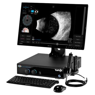
Escalon - Model VuMAX HD -Ophthalmic Ultrasound System
Unparalleled in every regard: image quality, advanced tools including automated quantitative angle analysis, sulcus-to-sulcus probe alignment, and more. Elegant, intuitive, exceptional.
Unparalleled Image Quality.
Hands down the gold standard in ophthalmic ultrasound. Unparalleled UBM and B-scan image quality with Enhanced Focus Rendering™ and large ultra high resolution screen allows you to capture both crisp still images and record video that can be carefully reviewed frame-by-frame.
Elegant user interface provides useful tools that are intuitive, simple, and efficient to use. Time-saving features such as selectable patient database display to easily search and access archive exam records. Document scan orientation with the single click of a button. Replay videos in real-time, slow motion, or frame-by-frame. Super-impose A-scan trace, perform linear and angle measurements, and annotate onto B-scan and UBM images. Auto calculation of axial length average and standard deviation, nine IOL formulas, and lens database for biometric A-scan.
Elegant user interface provides useful tools that are intuitive, simple, and efficient to use. Time-saving features such as selectable patient database display to easily search and access archive exam records. Document scan orientation with the single click of a button. Replay videos in real-time, slow motion, or frame-by-frame. Super-impose A-scan trace, perform linear and angle measurements, and annotate onto B-scan and UBM images. Auto calculation of axial length average and standard deviation, nine IOL formulas, and lens database for biometric A-scan.
ANTERIOR SEGMENT
View the entire anterior segment in great detail, with optimized scan settings of the VuMAX™ HD. Clearly visualize the ciliary body, pars plana, and other structures and identify tumors, cysts, trauma, uveitis, and other pathologies. The VuMAX™ HD even allows for visualization and video capture of accommodation.
ICL SIZING
Accurately measure sulcus-to-sulcus for properly and confidently size ICLs to ensure no post-operative surprises. The specialized sulcus-to-sulcus scan setting is optimally engineered to enable consistent viewing of key anatomical landmarks required to ensure accurate sulcus-to-sulcus measurements.
GLAUCOMA MANAGEMENT
The specialized angle detail scan setting optimizes resolution of different structures at the angle and behind the iris, providing the premier diagnostic tool for identifying causes of glaucoma-related concerns, including angle detail and permeability of the trabecular meshwork, plateau iris syndrome, effects of pupil movement on the angle, and other causes of glaucoma.
Unparalleled
Unparalled B-Scan.
The all-new B-scan mode of the VuMAX™ HD produces truly outstanding imaging of the posterior segment that has become the new gold standard.
SEE EXTRAORDINARY DETAIL WITH ENHANCED FOCUS RENDERING
Easily visualize extraordinary fine details within video clips and still images generated using proprietary Enhanced Focus Rendering™, providing image quality unmatched by other imaging systems.
OPTIMIZED ULTRASOUND IMAGING SETTINGS
MADE EASY
Select from 4 preset scan modes to optimize image quality in area of interest, including orbit, vitreous body, retina surface, and deep retina / choroid.
Quantitative Angle Analysis.
Accurately and consistently measure key parameters of the angle using the VuMAX HD UBM angle analysis tool. Easily track structure properties over time and assess differences during mydriatic and miotic conditions.
Sulcus-to-Sulcus Probe Alignment.
Measuring sulcus-to-sulcus is imperative for proper ICL sizing. The key to accurately measuring sulcus-to-sulcus is ensuring proper probe alignment. The VuMAX HD eye tracking feature provides real-time feedback to ensure your scans are properly aligned.