

- Home
- Companies
- Shared Imaging
- Products
- Shared Imaging - PET/CT Imaging Systems
Shared Imaging - PET/CT Imaging Systems
Shared Imaging provides PET/CT technology to hospitals, imaging centers, ambulatory/ED’s, and clinics. The company offers customizable solutions with technology options from various OEMs, allowing for tailored software packages, accessories such as injectors, and slice count configurations. PET/CT scanners integrate positron emission tomography (PET) with computed tomography (CT), enabling the combination of anatomical data from CT with molecular imaging from PET. This fusion facilitates the precise detection and mapping of abnormal cell functions. The PET component uses positron-emitting radionuclides to label biologically significant molecules, like sugars. The decay of these tracers is detected by the PET scanner, which creates physiologic maps of relevant functions or processes. Clinically, FDG, a glucose analog, is commonly used to image glucose metabolic rates in oncologic studies, exploiting the higher glycolytic rates of malignant cells. Shared Imaging also provides mobile imaging solutions, maintaining a premium fleet of medical coaches with ongoing maintenance to ensure high-quality service delivery.
Shared Imaging provides PET/CT technology to hospitals, imaging centers, ambulatory/ ED’s and clinics. With the choice of technology from most OEMs, we can customize software package, accessories (i.e. injectors) and slice count to match your clinical needs.
PET/CT scanners combine technology from two imaging modalities: positron emission tomography (PET) and computed tomography (CT). This combination makes it possible to fuse anatomic information from the CT scan with molecular imaging information provided by PET imaging. With this technology, not only can abnormal cell function be detected, it can be anatomically mapped with great precision.
The basis of the PET imaging component is the labeling of small, biologically important molecules, such as sugars, with positron-emitting radionuclides. When these positron-emitting tracers undergo radioactive decay, their positions can be detected by the PET scanner. By imaging the temporal distribution of these labeled compounds, “physiologic maps” of the functions or processes relevant to the labeled molecules can be created. Numerous different types of tracers have been developed for imaging with PET, but the vast majority of clinical oncologic PET studies performed at present utilize an analog of glucose, 18F-2-fluoro-2-deoxy-d-glucose (FDG). The use of FDG to image glucose metabolic rate takes advantage of the observation that malignant cells have higher rates of aerobic glycolysis than normal tissues (1). Thus, the malignant cell utilizes more glucose to meet its energy needs. For the typical clinical oncology study, FDG is administered intravenously in the quiet, resting state and is allowed to circulate through the body for 60 to 90 minutes before imaging is begun. In the case of most malignant neoplasms, sites of active tumor will show up as foci of hypermetabolism, or “hot spots” on the subsequent PET scan images.
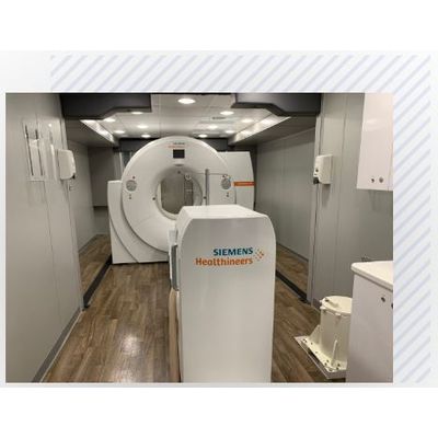
With the high demand for convenient access to care and the expectation of high-quality services by consumers – Shared Imaging treats their mobile medical coach fleet as premium medical technology. Shared Imaging practices proactive responsibility for on-going maintenance, repair, and maintaining the aesthetics of the interior and exterior of mobile medical coaches to ensure each coach and customer can deliver the quality of care that patients deserve.
- No build out required
- No large capital investment required
- Avoid space limitations
- Reduce patient backlog
- Relocatable for deployment at multiple sites
- Reduce operating costs while adding new or expanding existing service lines with upgraded or new imaging technology
- Provides convenient patient access
- Serve patients during construction or renovation, during equipment upgrade & downtime, or to recover quickly from natural disasters