

- Home
- Companies
- Mediso Ltd.
- Products
- nanoScan - Model SPECT/CT - Preclinical ...
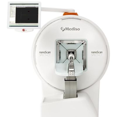
nanoScan - Model SPECT/CT - Preclinical Imaging Systems
Versatile SPECT/CT with absolute quantification and full stationary dynamic imaging. The nanoScan SPECT system images radiotracers with absolute quantification, high sensitivity and high spatial resolution down to 0.3 mm in vivo. Wide range of patented multi-pinhole collimators available with optimized performance for any applications: from whole-body to focused scanning; imaging of mice, multiple mice, large rats and rabbits; from low energy tracers to theranostic isotopes. Best homogeneity on the market is achieved by helical scanning, while fastest 3D dynamic acquisitions are carried out in full stationary mode without gantry or table motion. The SPECT system is also available in triple-modality combination with our high-end PET systems (see nanoScan SPECT/CT/PET).
- Proprietary and patented multi-pinhole collimator technology offering excellent spatial resolution, high sensitivity and large field of view at the same time
- Cutting-edge sensitivity even for high-energy isotopes ensured by 9.5 mm thick sodium iodide (NaI:Tl) crystals with large area and minimal detector gaps
- Exclusive imaging performance with ultra-low activity
- Tera-Tomo™ 3D SPECT reconstruction: iterative SPECT reconstruction engine based on real-time Monte Carlo simulation
- Full range of advanced corrections
- CT- or MRI-based attenuation and scatter correction
- Excellent homogeneity by helical scanning for comparison of regional uptakes
- Time-Activity Curve (TAC) generation and calculation of Standardized Uptake Values (SUV)
- PET-like dynamic imaging capability with list mode data storage
- Completely motionless acquisition providing time-continuous data from the whole cross-section of the animal
- 3D quantitative dynamic imaging with 1-3 second time frames
- Time-Activity Curve generation from 3D quantitative results.
- Large detector field of view:
27 cm x 27 cm (10.6” x 10.6”) - Wide gantry opening: up to 27 cm
- Various animal models from tiny mouse to large rabbits (6.5 kg / 14.3 lb)
- Multiple mouse imaging chamber with physiological monitoring feature for all animals
- Wide range of collimation techniques and collimators: Single-Pinhole, Multi-Pinhole and Parallel-hole.
- Dedicated multi-pinhole collimators for high-energy imaging
- List-mode data acquisition for simultaneous imaging of multiple isotopes, even for simultaneous scanning of PET and SPECT tracers
- 9.5 mm thick NaI:Tl crystals ensuring high sensitivity for high-energy gamma photons
- Imaging of theranostic isotopes like 131I, 213Bi, 225Ac and more
- Up to 80 W X-ray power enables high-performance scanning even for large or multiple animals
- Variable magnification for high-resolution imaging with 10 μm isotropic voxel size
- Very high throughput: fast scanning with real-time image reconstruction combined with multiple animal imaging capability
- Iterative image reconstruction for low-noise and low-dose imaging
- Ultra-low-dose protocol for follow-up studies with only 1 mSv whole-body dose to the animal
- Imaging with cardiac and respiratory gating
SPECT
up to 27 cm
mouse, rat, marmoset, guinea pig, rabbit (up to 6.5kg), monkey
up to 3 x 60 g mice
9.5 mm NaI(Tl)
down to 0.3 mm
up to 13 000 cps/MBq
from 125I to theranostic and PET isotopes
Multi-pinhole by proprietary M3-pinhole™ technologySingle-pinholeParallel-hole
Yes, ultra-fast 4D/5D list mode dynamic imaging by static or rotational SPECT
Acquisition schemes
SPECT (3D): helical, circular, semi-stationary and full stationaryPlanar (2D): static, dynamic
CT
up to 80 W
16 cm
12 cm
10 cm
mouse, rat, marmoset, guinea pig, rabbit
up to 3 x 60 g mice
modified Feldkamp-type for real-time reconstruction,iterative reconstruction for low-dose and low-noise applications
30 μm at 10 μm voxel size
Low-dose protocol
down to 1 mSv for whole-body mouse
MRI
Maintenance-free permanent magnet with 1 Tesla field strength
Zero (5 Gauss line is inside the cover)
Integrated RF shielding, no need for Faraday cage
Volume coils: whole-body mouse, whole-body rat Surface coils: mouse brain, rat brain
100 μm in vivo