

Neurescence - Model Chromatone -Solution for In Vivo Calcium Imaging and Optogenetics
Neurescence’s flagship product – The Chromatone system – provides a powerful integrated approach to observe, manipulate, and dissect a spectrum of neuronal subtypes in multiple brain/spinal cord regions, with single cell resolution. This three-pronged strategy to observe and stimulate activity in defined neuronal subpopulations, while examining behavioral paradigms will reveal accurate underlying mechanisms of physiology and behaviour in awake, freely behaving animals. The Chromatone system is the only complete solution for in vivo calcium imaging and optogenetics, that enables simultaneous multicolor imaging and multi-opsin stimulation in up to four brain or spinal cord areas in freely behaving animals.
Services included:
- Hands-on-surgical training
- Neuronal trace extraction support
- Device synchronization with other behavior equipment
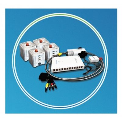
The components include the main microscope body, four illumination cubes, control electronics, multimode fibres and imaging fibre bundles. The microscope body is small and light weight, to ensure ease of use during a wide array of behavior paradigms and it integrates cell-type specific calcium imaging and optogenetics.
The illumination cubes contain excitation light sources and optical filters to ensure the correct illumination color arrives at the brain tissue. Active temperature control in the illumination cubes ensures highly stable illumination levels at the brain tissue, throughout a behavior session.
Each illumination cube connects to the microscope body through a multimode fiber and emits three illumination colors:
- 470nm +/- 12nm for Green Fluorescent Protein Imaging /ChannelRhodopsin optogenetics
- 550nm +/-7.5nm for Red Fluorescent Protein imaging
- 632nm +/- 14nm for ChrimsonR Optogenetics
Light from each illumination cube is coupled into an imaging fiber bundle, which carries the light to implanted GRIN lens connectors and then to the brain tissue.
Video 1: GFP imaging ChrismsonR stimulation;
Video 2: GFP & RFP imaging ChrismsonR stimulation;
Video 3: RFP imaging ChannelRhodopsin stimulation;
Video 4: RFP imaging ChrimsonR stimulation
There are four such imaging fiber bundles, enabling simultaneous multicolor calcium imaging and multi opsin stimulation in up to four brain or spinal cord areas.
Pictured above is a depicted behavior session using the ChromatoneTM system.
During an imaging -behavior session, the imaging fibre bundles of the ChromatoneTM are connected to the implanted lens-connectors. Three different illumination colors are available in each implanted region, to enable multicolor imaging (GFP and/or RFP) and multi-opsin optogenetics (ChannelRhodopsin / ChrimsonR). The three illumination colors red, blue and green in each implanted region can be independently controlled using the software.