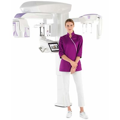


Model Hyperion X9 Pro - The 3-in-1 System
- Easily upgraded to all configurations
- Reversible CEPH arm
- Operates with relocatable 2D sensor or two sensors
- The most compact 3-in-1 system
- Direct conversion 2D sensor
DCIII technology applies the innovative direct conversion sensor that has revolutionised PiE (Powerful image Enhancer) 2D imaging. Standard systems convert X-rays into visible light which is, in turn, converted into electrical signals to create the digital image. With DCIII technology, instead, the sensor receives and processes the X-rays directly, resulting in increased sensitivity and efficiency without any loss of detail. This lets users obtain both high resolution images with greater contrast at low doses and extremely detailed images from fast-scan, ultra-low dose protocols such as QuickCEPH or QuickPAN.
- Multi FOV from 4 x 4 to 13 x 16 cm
- Upgraded generator
- Extremely high resolution (up to 68μm)
- Fast CB3D scan (as brief as 3.6 s)
- Low dose
Ear: 7 x 6 cm (XF)
Nose and maxillary sinuses: 13 x 8 cm
Mouth and Throat: 13 x 10 cm
Complete upper airways: 13 x 16 cm
ADVANCED
Dentition up to frontal sinuses: 13 x 16 cm
Ascending mandibular branches: 13 x 10 cm
Zygomatic arches and sinuses: 13 x 8 cm
Maxillary sinuses: 10 x 10 cm
Teeth: 4 x 4 cm (XF)
BASIC
Complete dentition, adult: 10 x 8 cm
Single dental arch, adult: 10 x 6 cm
Complete dentition, child: 8 x 8 cm
Single dental arch, child: 8 x 6 cmHemiarch or anterior dentition: 6 x 6 cm
TMJ: 7 x 6 cm (XF) open mouth/closed mouth
Cervical spine: 9 x 9 cm (XF) - Voxel 68 μm
DOUBLE DENTAL ARCH SCAN AT 75 μm
FOV with a 10 cm diameter, also essential for reliable acquisition of the complete roots of impacted third molars and height up to 10 cm. At an exceptional resolution of 75μm, Hyperion X9 pro provides, with a single acquisition, images of the entire dentition and the surrounding bone structures. The perfect tool to plan multiple implants, also with the use of surgical guides.
FULL AIR WAYS
The 13 x 16 cm FOV captures the complete upper airways in one single examination. Detailed view of the entire dentition, maxillary sinuses and upper airways, so as to clearly identify possible signs of narrowing and correctly diagnose obstructive sleep apnea syndromes (OSAS).
