

- Home
- Companies
- Ionpath, Inc.
- Products
- MIBIscope - Transforming Multiplexed ...
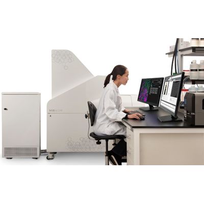
MIBIscope - Transforming Multiplexed Tissue Imaging System
The MIBIscope System is a revolutionary imaging platform, enabling comprehensive phenotypic profiling and spatial analysis of the tissue microenvironment. The MIBIscope allows researchers to visualize over 40 markers simultaneously with higher sensitivity, resolution, and throughput than existing methods.
- Visualize 40+ markers in a single scan
- Comprehensively phenotype immune infiltrate
- Quantify protein expression
- Profile tissue architecture
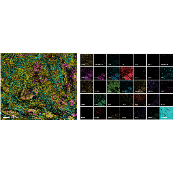
Visualize 40+ Markers in a Single Image
The MIBIscope enables over 40 markers to be visualized simultaneously with single step staining and single step imaging.
MIBIscope images from a 3mm x 3mm scan of a granulomatous lung section from a Mycoplasm tuberculosis infected patient. The image to the left shows immune infiltrate in the infected tissue (CD45, yellow; CD31, red; SMA, blue; CD68, magenta). Zoomed in single-channel images shown to the right.
- Available Biomarker Channels: 40
- Resolution: 350 nm – 1 µm
- ROI Size: 400x400 – 800x800 µm2
- Acquisition Time (per 800x800 µm2):
- Coarse Resolution (1 µm), 35 min
- Fine Resolution (500 nm), 68 min
- Super-fine Resolution (350 nm), 139 min
- Lower Limit of Detection (number of Ab): 1 (113In) – 16 (166Er)
- Dynamic Range: 5 log
- File Type: TIFF