

Ultrasound
Ultrasound is the most widely used and a cost-effective imaging technology. The combination of ultrasound contrast agents with the latest evolving equipment has the potential to transform the clinical role of ultrasound to provide a flexible, cost-effective imaging capability equivalent to existing established modalities such as MRI, CT, and X-ray.
Ultrasound scanning is patient-friendly and doesn’t expose the patient to radiation, unlike X-ray imaging. Ultrasound contrast agents don’t cause damage to the kidneys and are rapidly eliminated allowing the healthcare personnel to perform repeated examinations in short intervals.
In echocardiography (cardiac imaging), the standard examination for assessment of cardiac anatomy and function, the endocardial border is sometimes difficult to recognize due to limited image resolution. This is particularly an issue when treating obese patients. Embedding an ultrasound agent in the procedure can result in a significant improvement in image quality and therefore the treatment1.
Doppler ultrasound is the standard examination technique for assessment of vascular pathologies. However, the method is very angle dependent and only able to detect flow towards or away from the transducer causing limitations in case of slow velocities or small volumes. CEUS has a much higher sensitivity, allowing detection low concentration of microbubbles in the blood even at zero velocity.
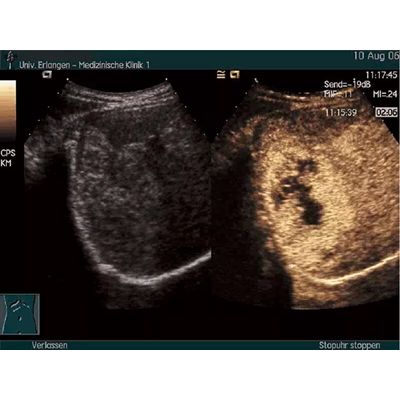
In many cases, small vessels inside parenchymal lesions cannot be assessed. The use of ultrasound agents allows the depiction of even small and irregular vessels – and more importantly, the assessment of the enhancement pattern based on the intensity of the enhancement, the time of the contrast arrival, and the detection of wash-out. This is the basis for the characterization of focal lesions in the liver and breast2.
Urine reflux from the urinary bladder upwards to the kidney in children is a common reason for recurrent urinary tract infections – which can result in irreversible kidney damage. A standard imaging test has been an X-ray examination with intravesical administration of an iodine contrast agent for assessment of the excretory urinary tract. Using an ultrasound contrast agent instead, a similar examination can be performed without ionizing radiation4,5.