

X-ray Imaging
Halifax Biomedical Inc. is developing Stereo Orthopaedic Radiography (SOR) technology that provides solutions to two of the largest problems in orthopaedics: instability of the spine, and loosening of implants such as hip and knee replacements. Both problems are difficult to detect and measure objectively and accurately in three dimensions.
The spine has two primary functions: to protect the spinal cord and to allow movement. These competing functions create a delicate balance between mobility and stability causing even very subtle excessive motions to possibly create clinical symptoms. Halifax Biomedical Inc. is developing a technology that will enable the measurement of these subtle excessive motions.
Much in the way houses shift and settle over time, implants do the same thing after initial implantation. This is common and it isn’t always a problem, but sometimes it can lead to patients experiencing pain or other symptoms that need to be treated. Stereo radiography allows surgeons to identify very subtle movements between tests and monitor these over time.
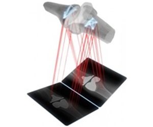
Stereo Orthopaedic Radiography (SOR) (also known as Roentgen Stereophotogrammetric Analysis or RSA) is a methodology which uses two x-ray images taken at the same time for the purpose of making accurate 3D measurements. Much like having two eyes vs. one, SOR allows for accurate depth perception.
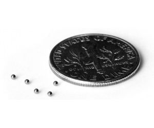
There are several ways to perform SOR. The traditional and most accurate method involves implanting tiny markers (tantalum balls of 1.0 mm diameter) into a patient’s bone during surgery and attaching these markers to an implant. This allows for accurate measurement of the motion of the implant relative to the bone via the markers. Early excessive motion has been shown to predict implant loosening later on. Wear of the implant’s insert can be measured with this methodology as well.
Implants can also be measured directly eliminating the need to attach markers which can be a costly endeavor due to regulatory requirements. Direct measurement is only possible if accurate CAD models or reverse engineered models are available. This results in a small but acceptable loss in accuracy for most applications.
Both previous methods require a surgical procedure. For pre-operative measurements no implants of markers are available to perform the measurements. This is the most challenging form of SOR, because there is less contrast between bones and soft tissue and there is no prior knowledge of the exact shape of the bone. Shape models, however, can be reconstructed from 3D imaging data such as CT and MRI, but this is a prohibitively labor-intensive method. Halifax Biomedical Inc. is developing a novel technique that will estimate the shape of the bone, based on knowledge of the anatomy of that bone, and the bone’s position and orientation in the same analysis eliminating the need for 3D imaging.
Halifax Biomedical Inc. has developed a complete Stereo Orthopaedic Radiography (SOR) solution from the operating room, to imaging, to the doctor’s office by providing purpose-built surgical tools and imaging equipment as well as stability assessment services. This provides a chain of processes optimized for ease of use, consistency and accuracy of the data, and minimal impact on work flow.
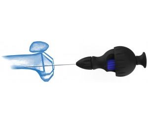
While performing surgery, the surgeon uses the Halifax Bead Inserter to place tiny beads in the bone to act as reference points. A minimum of three, and preferably five or six, of these beads are placed in optimized locations so that they are visible from multiple views. This straight-forward procedure takes 2 to 3 minutes on average and enables surgeons to gather vast amounts of data about a patient down the road. Beads can be placed in almost any bone in the body for monitoring and diagnostic purposes.
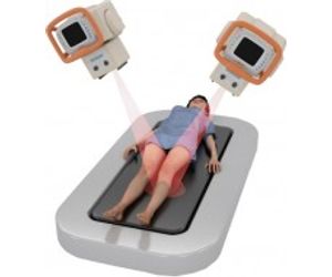
Patients are brought back to the clinic for regular follow-up imaging after their surgery. Unlike conventional x-ray, low-dose SOR images are taken at two different angles allowing for three-dimensional stability assessment of the patient’s bone and implants. Halifax Biomedical Inc.’s Halifax Imaging Suite ( or Kit) options use cutting-edge digital imaging equipment to increase precision of measurements while at the same time reducing patient dose. SOR images are transferred to Halifax Biomedical Inc. in an anonymized way so that patient confidentiality is guaranteed.
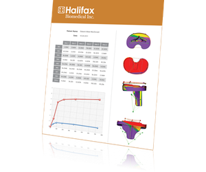
The images are analyzed by expert analysts and the very precise measurements are sent back to the surgeon for interpretation in the form of a detailed report. This stability assessment can be done on hip and knee replacements, as well as ankle, spine, and many other joints, and is performed using Model-Based RSA (RSAcore, Department of Orthopaedics, Leiden University Medical Center, Leiden, The Netherlands). Pre-operative spine measurements are performed with Halifax Biomedical Inc.’s proprietary software.
