

- Home
- Companies
- Indica Labs Inc.
- Software
- Halo - Version AI - Deep Learning ...
Halo - Version AI -Deep Learning Classifier Add-on Software
HALO AI is a collection of train-by-example classification and segmentation tools underpinned by advanced deep learning neural network algorithms. HALO AI classifiers can be trained to quantify tissue classes, to segment tissue classes for analysis with other HALO image analysis modules, to find rare events or cells in tissues, and to categorize cell populations into specific phenotypes.
SIMPLE & INTUITIVE WORKFLOW
HALO AI is fully integrated with the intuitive, easy-to-use HALO and HALO Link viewers and employs a simple three-step workflow. After defining what tissue classes or cell phenotypes you would like to segment, you train the neural network by drawing annotations – no computer programming or AI knowledge required. Trained classifiers can be applied to segment tissue and cells on any whole slide image or region of interest.
POWERFUL TISSUE SEGMENTATION
HALO AI now includes the option of three powerful neural networks – VGG, DenseNet and MiniNet. VGG, a well known and more traditional network, was used to build the Indica Labs submission in the CAMELYON17 challenge and was the first neural network integrated with HALO AI. DenseNet is a more modern network capable of creating more robust classifiers at higher resolution compared to VGG. MiniNet, a custom network developed at Indica Labs, is more shallow than VGG or DenseNet, but can produce a solution quickly with limited training data and is therefore useful for testing new AI applications. Skip down to see some applications tested with our built-in networks.
EXCEPTIONAL CELL CLASSIFICATION
Segment nuclei with the nuclei Segmentation classifier. Utilize HALO AI’s pretrained networks for H&E, single IHC, or DAPI stained images for an out of the box solution. Or train your own nuclei segmentation network for a specific application (unique tissue or advanced staining protocols). Once nuclei are segmented, take it a step further using the Nuclei Phenotyper classifier to automatically assign cells into user defined phenotypes with a few quick training examples. Keep scrolling to see some applications tested with our Nuclei Segmentor and Nuclei Phenotyper.
The HALO AI VGG network was employed to detect and stage metastatic tumor cells in H&E-stained lymph nodes of breast cancer patients in this example. While the VGG network requires more data and training, it works well on examples like this where the stain intensity and morphology are variable. Download the application note describing the Indica Labs’ submission to the CAMELYON17 project.
The evaluation and quantification of islets in H&E-stained tissue sections can be very time-consuming due to heterogeneous morphology of islets. For this application, we train the VGG network to identify islets within human pancreatic tissue sections. Download the application note describing the method used to build a classifier for accurately segmentation of islets from surrounding exocrine tissue, irrespective of stain or morphological variability.
For this application, MiniNet was used to separate tumor from lymphocytic clusters and stroma in and H&E stained breast cancer tissue. MiniNet is a shallow neural network, but it works well on routine classification tasks and may be used as an alternative to HALO tissue classifier (random forest) in some instances. It is also recommended for quick proof-of-concept studies before moving over to the more data and time intensive VGG and DenseNet networks. Open full-sized image.
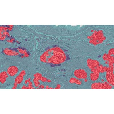
Here the HALO AI DenseNet neural network was used to segment out glomeruli in differently stained kidney sections, including Jones’ silver stain, H&E, ISH and IHC. In the silver stained slide, the glomeruli and tubules are highly variable in color and morphology. In the ISH, IHC and H&E-stained sections, there is little to no color contrast between the cells in the glomeruli and the tubules. Open full-sized images.
While the masking networks (MiniNet, DenseNet and VGG) can be trained to segment tissue classes, they cannot be applied to classify individual cells. For this application, we use the Nuclei Phenotyper. For this application, nuclei are first segmented using a pre-trained Nuclei Segmentation network and then the individual nuclei are phenotyped using training examples provided by the pathologist (shown here, tumor cells in blue, stroma cells in green and lymphoctyes in yellow).