

- Home
- Companies
- ClearLight Biotechnologies, Inc.
- Training
- Tissue Clearing Primer
Tissue Clearing Primer
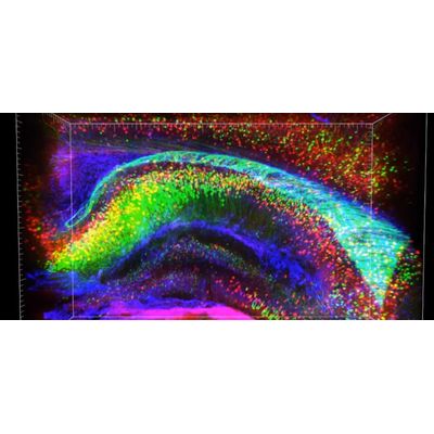
CLARITY is an acronym for Clear Lipid-exchanged Acrylamide-hybridized Rigid Imaging / Immunostaining / in situ-hybridization-compatible Tissue hYdrogel). Discussions about CLARITY tissue clearing inevitably involve imaging. The two go hand-in-hand. Tissue clearing is what you need to do in order to see beyond the tissue surface at a cellular and subcellular level. It is akin to being able to see in the darkness using night vision goggles or using sunglasses to be able to clearly see the landscape in the presence of bright sunlight. When researchers and scientists want to see inside of a brain, or tumor or study diseased or healthy tissue they first will want to have that tissue cleared of the things that occlude the view.
Most tissue is opaque, meaning that visible light cannot travel through it. For example, you can’t see through your hand. If you hold your hand up to a light source, it will block the light and cast a shadow. If you fall and injure your hand, your doctor can use an x-ray machine to see if you may have broken a bone.
Unlike visible light, x-rays pass easily through the soft tissue of your hand, like skin, muscle and tendons, but they scatter when they encounter dense material such as bone. Because of this property, x-rays are ideal for imaging broken bones and discovering tooth decay.
To study structures in soft tissue—especially very small, delicate structures, like neurons in a brain, or cancerous lesions in breast tissue, scientists need a way to see beyond the tissue surface.
Historically, this meant putting thin slices of tissue onto glass slides and then staining and examining the slides one by one under a microscope. They could only guess how the tiny tissue structures actually looked and functioned in the whole sample before being dissected and destroyed in the process of analysis.
These techniques are over 100 years old, and yet still represent the current gold standard. Scientists and researchers still primarily rely on the analysis of 2D thin section FFPE (~5-10 micron) samples. Using these methods, spatial and morphological analysis of the tissue microenvironment is constrained. Additional limitations include the tedious and labor intensive processing of hundreds of slides. Therefore, researchers using the current gold standard rely on a small sampling of the tissue in attempting to understand complex biological processes. This is the problem with 2D FFPE.
Although rudimentary types of tissue clearing have been around since the early 20th century, they were mostly useful for large samples. Small and subcellular details were destroyed in these early methods. In the last 5 - 10 years, modern tissue clearing techniques, especially those that can be combined with immunohistochemical staining techniques, are revolutionizing how we study subcellular tissue morphology.