Fonar Corporation
- Home
- Companies
- Fonar Corporation
- Articles
- Postoperative Spinal Instability at ...
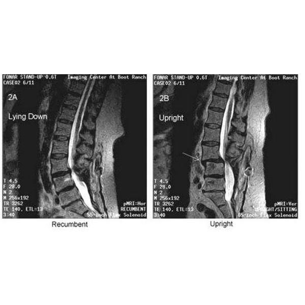
Postoperative Spinal Instability at L3-4 Revealed by Upright Weight-Bearing MRI - Case Study
Mar. 16, 2021
Courtesy ofFonar Corporation
The patient was scanned in the FONAR Upright MRI in early 2002, one year after her spinal fusion. Both Upright and recumbent scans were performed on her in the multi-position FONAR Upright MRI. The recumbent MRI (left image) exhibited only a normal lumbar lordotic curve and a modest bulge of the L3-4 intervertebral disc, consistent with her prior recumbent MRI scan on a conventional MRI. The FONAR Upright scan (right image) revealed, however, a marked subluxation (anterolisthesis) at L3-4 and an accompanying spinal stenosis that were not visible on the recumbent MRI.
Most popular related searches