

- Home
- Companies
- NDI Europe GmbH
- Products
- Aurora - Electromagnetic (EM) Tracking ...
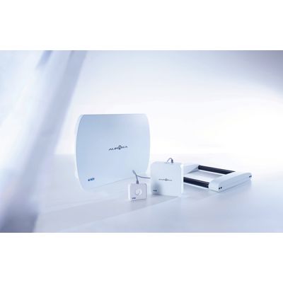
Aurora - Electromagnetic (EM) Tracking System
The Aurora® electromagnetic (EM) tracking system provides real-time unobstructed tracking of miniaturized sensors embedded into surgical tools, probes, needles, guidewires, and catheters. The position and orientation of each sensor is tracked in five or six degrees of freedom (5DOF / 6DOF), both inside and outside the body. No line of sight is needed to maintain sub-millimetre, sub-degree tracking. A sensor can even be placed at the tip of a flexible instrument, extending the benefits of 3D localization and guidance to countless new medical procedures. The Aurora will simultaneously track up to sixteen (16) 5DOF or eight (8) 6DOF tools within an EM-generated tracking volume. Different Field Generators (FG) provide optimal tracking volumes for a variety of applications. The result is highly accurate and reliable EM tracking required by the most precise image-guided surgery and interventional applications in medicine.

Field Generator
Emits a low-intensity, varying electromagnetic field and establishes the position of the tracking volume. NDI now offers multiple FG types that feature plug-and-play functionality with the System Control Unit.
Aurora® Planar Field Generator
For mounting on a positioning arm which offers flexible setup options around the patient.
Aurora® Tabletop Field Generator
Only 3.4 cm thick, this FG is designed to be placed between the patient and the table, and incorporates a thin barrier that minimizes tracking distortions caused by conductive or ferromagnetic materials located below it.
System Control Unit (SCU)
Controls the FG, collects information from the SIUs, calculates the position and orientation of each sensor, and interfaces with the host computer. Available in both enclosed and PCB formats.
Sensor Interface Unit (SIU)
Amplifies and digitizes the electrical signals from the sensors and provides an increased distance between the SCU and sensors, while minimizing the potential for data noise. Up to 2 SIUs can be connected. Available in both enclosed and PCB formats. Supports eight 5DOF or four 6DOF sensors.
Sensors and Tools
NDI offers a wide selection of both individual sensors and ready-to-use tools for use with the Aurora System.
OEM Customization
For over 30 years, NDI’s measurement technology solutions have been integrated into the world’s most sophisticated surgical navigation systems. The Aurora continues this tradition, bringing real-time electromagnetic tracking technology to a wide spectrum of image-guided surgery and interventional systems. Aurora components are customizable, allowing for the creation of new system form-factors, and seamless integration into existing workflows.
- Customized FGs are part of the system as opposed to external hardware that hinders workflow
- Customized electronic components allow for different system configurations
- Customized sensors and tools for unique tracking requirements
- Customized services including tool design, sub-tool assembly, and tool characterization
Applications
Interventional Radiology and Imaging
- Enable new ultrasound imaging capabilities through localization of the ultrasound transducer, and overlay live ultrasound images on pre-proceduraly acquired CT or MRI images through image fusion.
Oncology
- Track needle tips within fluoro, CT, ultrasound, or MRI images when performing core tissue biopsies, fluid aspirations, brachytherapy, and radiation therapy.
Guidewire and Catheter Tracking
- Track guidewires and catheters used in endovascular procedures such as renal denervation, stent and shunt placement, abdominal aortic aneurysm repair, and more.
Scope Tracking
- Track the absolute and relative poses of a laparoscopic ultrasound, the tip of a bronchoscope or pulmonary catheter, and the position and orientation of rigid and flexible endoscopes.
ENT Surgery
- Track surgical instruments to confirm and facilitate procedural steps, and to avoid cranial anatomy close to the sinuses.
Cardiology
- Create high-resolution spatial maps of electrical activity of the heart by tracking electrophysiology catheters.
Orthopaedic Trauma
- Target distal locking holes in intramedullary nails without the need for fluroroscopy.