

- Home
- Companies
- Centinel Spine
- Products
- Stalif - Model M Ti - No-Profile ...
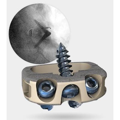
Stalif - Model M Ti -No-Profile Anterior Lumbar Integrated Interbody System
Sixth generation STALIF® technology, STALIF M-Ti™ incorporates a novel, circumferential, titanium surface, PEEK Integrated Interbody™ design that is raising the Gold Standard of lumbar interbody fusion.
Raising the Standard with
The Ti-ACTIVE™ surface provides a three-dimensional texturized surface that is microporous with hydrophilic properties, to facilitate human mesenchymal stem cell (hMSC) attachment and proliferation as demonstrated by pre-clinical testing performed on representative PEEK coupons with Ti-ACTIVE surface.2
During manufacturing, commercially pure titanium (CP Ti) is applied to the PEEK cage by a plasma spray to create our Ti-ACTIVE devices. The unique CP Ti surface system utilizes a robotic applicator, enabling the creation of a high-quality, reproducible titanium surface.3 Ti-ACTIVE was tested for mechanical integrity and the results exceeded the FDA requirements for shear and tensile strength for metallic spray surfaces.4
SEM image (2,000x magnification) of the surface of a standard PEEK implant (left) and a titanium-applied PEEK implant (right). Unlike standard PEEK, the PEEK with titanium surface provides a 3-D microporous environment conducive to cell attachment and cell migration, seen by cytoplasmic expansions of the cultured cells, as highlighted in purple.