

- Home
- Companies
- Newcells Biotech Limited
- Products
- Retinal Organoids
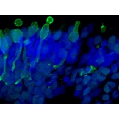
Retinal Organoids
Light responsive retinal organoids for accurate prediction of clinical outcomes. The retinal organoids recapitulate the complex structure of the human retina with laminar cell organisation mimicking embryonic development. They contain the outer photoreceptor segment of the retina that responds to light. Available analytical readouts: Immunofluorescence analyses, mRNA quantification by RT-qPCR, Transcriptomic analysis by single-cell RNA sequencing, Cytotoxicity assays, Cytokine release, Flow cytometry, Electron microscopy
- Fresh retinal organoids
- Retinal organoid cell pellets
- Total RNA preparation of retinal organoids
- Frozen sections of retinal organoids
- Available at various customisable time points e.g. D60, D150, D180, D210
- Rod and cone photoreceptors
- Retinal ganglion cells
- Bipolar cells
- Horizontal cells
- Amacrine cells
- Müller glial cells
- Human (Healthy donor)
- Human (Gene-edited)
Accelerate your lead compound selection by understanding their mode of action in functional retinal tissue
- Complex functional human retina model
- Well-characterised and reproducible
- Ready-to-use
Newcells provides on demand supply of fresh retinal organoids through regular batch release every 4-6 weeks after receipt of a purchase order. We also provide retinal organoids as cell pellets, RNA and frozen sections slides to suit multiple applications.
Take advantage of our expertise: with our fast and reliable service, we design the experiments with you to answer your questions and give you the results within 2 months. Our bespoke projects are carried out in our state-of-the-art UK facilities.