

- Home
- Companies
- Mimetas B.V.
- Products
- OrganoReady - Angiogenesis HUVEC
OrganoReady - Angiogenesis HUVEC
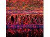
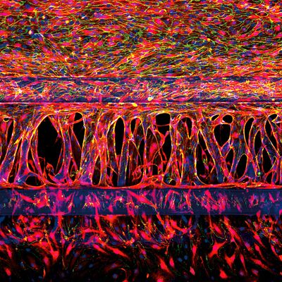
OrganoReady® Angiogenesis HUVEC comprises 64 HUVEC tubules ready-to-sprout. The product is based on the OrganoPlate® 3-lane 64 with 3 adjacent channels per chip, including one perfused HUVEC tubule. Do you want to perform experiments in human-relevant, complex 3D tissue models but don’t have the time or experience to build your own? Look no further, with the OrganoReady Angiogenesis HUVEC you can now take your angiogenesis research to the next level. Create and set up your desired studies such as compound profiling, screening or subsequently investigate sprouting behavior.
- OrganoPlate® 3-lane 64, including:
- 64 HUVEC tubules ready to sprout
- Sprout initiation mix: OrganoMedium HUVEC-AN-I
- Perfused 3D microvasculature
- Tip-stalk cell hierarchy & anastomosis
- HUVEC cell line: Human umbilical vein endothelial cells
- ECM: Collagen I (Rat Tail)
Each tissue culture chip contains one in-gel culture channel and two perfusion channels, of which one perfused HUVEC tubule. Compounds and stimuli can directly be added from the apical and basolateral sides of the culture. With this direct access, the platform enables you to investigate microvascular damage, tumor angiogenesis, fibrosis, and sprout formation.
Meet the OrganoReady Angiogenesis HUVEC
Ready-to-sprout blood vessel tubules
64 HUVEC tubules ready-to-sprout, seeded against collagen I, allowing you to perform assays right away.
We create the model, so you can focus on your assays
Preparation and QC of OrganoReady Angiogenesis HUVEC are done by our experts. Use the OrganoReady Angiogenesis HUVEC directly after arrival for up to 6 days.
Get inspired by a wide range of possible applications
Study for example (drug-induced) toxicity, compound profiling, use it for disease modeling or for fundamental research on angiogenic sprouting behavior.
No artificial membranes for free migration, invasion, and exchanges of factors
Unique PhaseGuide™ technology allows cells to interact and migrate freely between channels.
We have optimized the OrganoPlate® platform for growing perfusable 3D angiogenic sprouts, including tip-stalk cell hierarchy and anastomosis. The platform integrates both perfusion and the generation of stable biomolecular gradients and demonstrates its potential to study more physiologically relevant angiogenic sprouting and microvascular stabilization.
In this video, we show you in a nutshell how 3D angiogenesis models are developed at MIMETAS. Watch it now and learn how we can support your research.